|
||
|
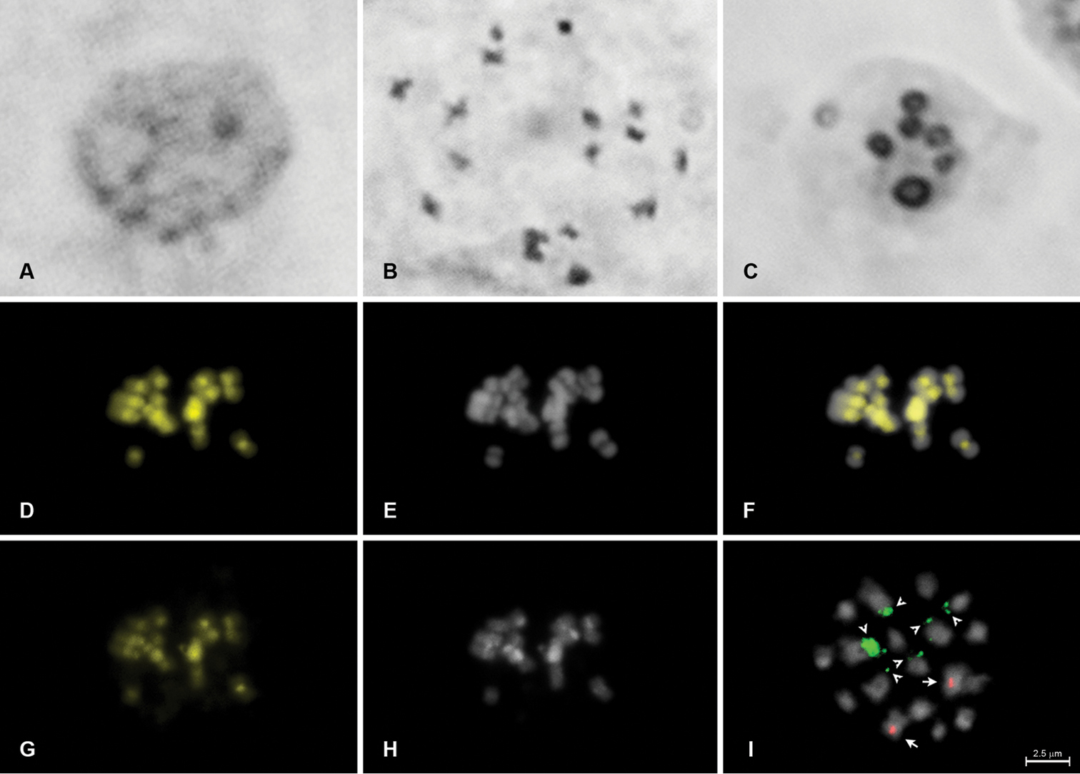
Karyotype analysis of Euphorbia hirta (2n = 18). Standard staining of mitotic interphase nucleus (A); standard staining of mitotic metaphase (B); silver impregnation of mitotic interphase nucleus (C); fluorochrome banding of metaphase chromosomes stained with CMA (D) and DAPI (E) and superposed images (F); C-banding of chromosomes stained with CMA/DAPI (C-CMA/DAPI; G–H); and metaphase chromosomes hybridized with 5S (red) and 45S (green) rDNA probes (I). Arrows and arrowheads indicate 5S and 45S rDNA sites, respectively. |