|
||
|
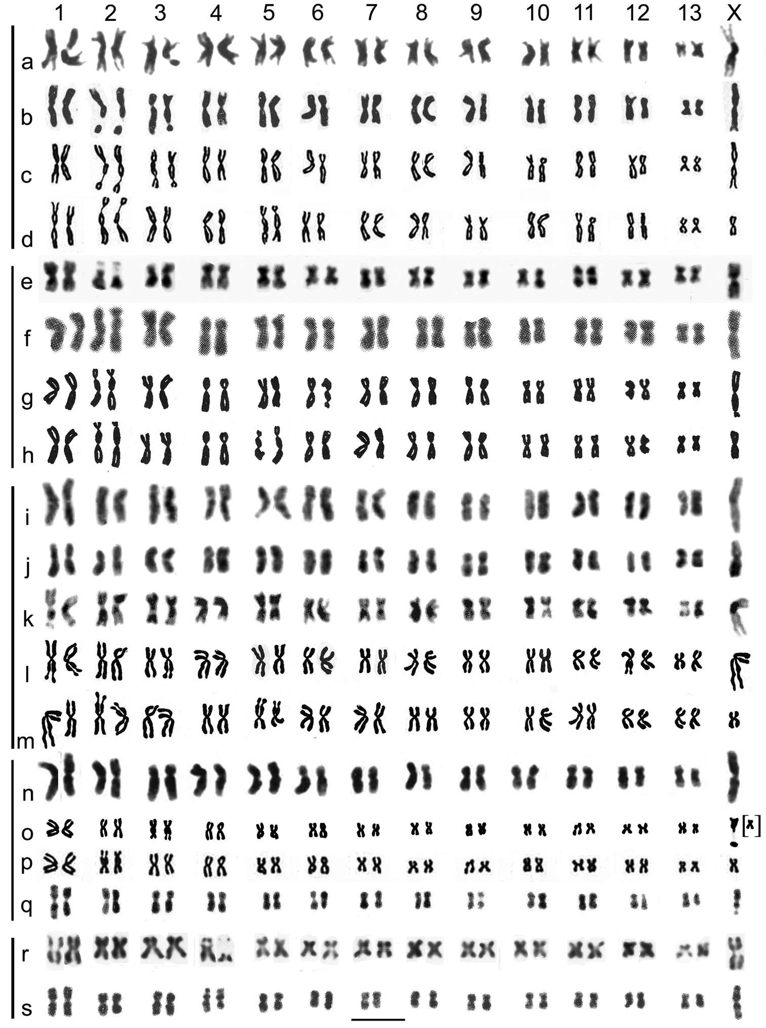
Mitotic chromosomes of Gyrinus spp, arranged as karyotypes, to compare the present results with those of Saxod and Tetart (1967) and Tetart and Saxod (1968). a–d G. caspius, a present material (Fig. 5b) b Saxod, Tetart, photograph (Plate 1B) c idem, drawing (Fig. 2), arranged as Fig. 5b, d the same drawing as arranged by Saxod, Tetart e–h G. paykulli, e present material (Fig. 5c) fTetart and Saxod (1968), photograph (Plate1A) g idem, drawing, (Fig. 1), arranged as e, h the same drawing as arranged by Tetart, Saxod i, j G. distinctus fairmairei, present material (Fig. 5f, g) k–m G. distinctus distinctus from Saxod and Tetart (1967)k photograph (Plate 1A) l idem, drawing (Fig. 1), arranged as the present G. d. fairmairei (i, j) m the same drawing as arranged by Saxod, Tetart n–q G. substriatus n present material (Fig. 5q) o drawing by Tetart, Saxod (Fig. 2) but with the X chromosome taken from their photograph (idem, Plate 1C), arranged as present material (n) p the same drawing as arranged by Tetart, Saxod. The partial X chromosome is placed as the right-hand replicate of chromosome 13 q karyotype prepared from Saxod, Tetart (Plate 1D) r, s G. suffriani r present material (Fig. 5t) s karyotype prepared from Saxod and Tetart 1967 (Plate 1C). The horizontal scale-line represents 5 μm. The vertical lines on the left hand side link karyotypes of the same species. |