Comparative Cytogenetisc 5(3): 211–221, doi: 10.3897/CompCytogen.v5i3.1435
Genomic and karyotypic variation in Drosophila parasitoids (Hymenoptera, Cynipoidea, Figitidae)
Vladimir E. Gokhman1, J. Spencer Johnston2, Chiyedza Small3,4, Roma Rajwani3, Shawn J. Hanrahan2, Shubha Govind3,4
1 Botanical Garden, Moscow State University, Moscow 119991, Russia
2 Department of Entomology, Texas A&M University, College Station, TX 77843-2475, USA
3 The City College of The City University of New York, Biology Department MR526, 138th Street and Convent Avenue, New York, NY 10031, USA
4 The Graduate Center of The City University of New York, 365 Fifth Avenue, New York, NY 10016, USA

Corresponding authors: Vladimir E. Gokhman (gokhman@bg.msu.ru), J. Spencer Johnston (spencerj@tamu.edu), Shubha Govind (sgovind@ccny.cuny.edu).

Academic editor: Seppo Nokkala

received 13 May 2011 | accepted 16 June 2011 | Published 24 August 2011


(C) 2011 Vladimir E. Gokhman. This is an open access article distributed under the terms of the Creative Commons Attribution License, which permits unrestricted use, distribution, and reproduction in any medium, provided the original author and source are credited.


For reference, use of the paginated PDF or printed version of this article is recommended.

Abstract

Drosophila melanogaster Meigen, 1830 has served as a model insect for over a century. Sequencing of the 11 additional Drosophila Fallen, 1823 species marks substantial progress in comparative genomics of this genus. By comparison, practically nothing is known about the genome size or genome sequences of parasitic wasps of Drosophila. Here, we present the first comparative analysis of genome size and karyotype structures of Drosophila parasitoids of the Leptopilina Förster, 1869 and Ganaspis Förster, 1869 species. The gametic genome size of Ganaspis xanthopoda (Ashmead, 1896) is larger than those of the three Leptopilina species studied. The genome sizes of all parasitic wasps studied here are also larger than those known for all Drosophila species. Surprisingly, genome sizes of these Drosophila parasitoids exceed the average value known for all previously studied Hymenoptera. The haploid chromosome number of both Leptopilina heterotoma (Thomson, 1862) and Leptopilina victoriae Nordlander, 1980 is ten. A chromosomal fusion appears to have produced a distinct karyotype for Leptopilina boulardi (Barbotin, Carton et Keiner-Pillault, 1979)(n = 9), whose genome size is smaller than that of wasps of the Leptopilina heterotoma clade. Like Leptopilina boulardi, the haploid chromosome number for Ganaspis xanthopoda is also nine. Our studies reveal a positive, but non linear, correlation between the genome size and total chromosome length in Drosophila parasitoids. These Drosophila parasitoids differ widely in their host range, and utilize different infection strategies to overcome host defense. Their comparative genomics, in relation to their exceptionally well-characterized hosts, will prove to be valuable for understanding the molecular basis of the host-parasite arms race and how such mechanisms shape the genetic structures of insectcommunities.

Keywords

Drosophila, Figitidae, parasitoid, genome size, karyotype

Introduction

Each species has a characteristic genome size and chromosome number. This information often serves as a starting point for obtaining whole genome sequence. It is also useful for cytological or PCR-based genotyping and comparative genomics. Drosophila melanogaster Meigen, 1830 is by far the best-studied insect. Availability of its annotated sequence data (Flybase 2011) is facilitating rapid progress as details of novel gene functions are uncovered and analysis of gene interaction networks and pathways is becoming possible. Sequencing of the Drosophila melanogaster genome also provided the baseline for the analysis of eleven additional Drosophila Fallen, 1823 species, spurring detailed investigation of the evolution of biological processes (Crosby et al. 2007).

Many species of Drosophila serve as hosts to parasitic wasps (Schlenke et al. 2007). In spite of spectacular progress on the model organism itself, practically nothing is known about the genomics or genetics of the parasitic wasps. Leptopilina Förster, 1869 and Ganaspis Förster, 1869 species (Figitidae) attack larval stages, emerge as free-living adults from the pupal cases of their hosts (Schilthuizen et al. 1998, Melk and Govind 1999, Allemand et al. 2002). Leptopilina boulardi (Barbotin, Carton et Keiner-Pillault, 1979) is a specialist parasitoid, while Leptopilina heterotoma (Thomson, 1862) is a generalist; these species exhibit distinct strategies to evade or overcome host defense (Schlenke et al. 2007, Kraaijeveld and Godfray 2009, Lee et al. 2009). Drosophila-Leptopilina interactions have increasingly become important in understanding innate immunity against natural metazoan parasites and the molecular basis of the arms race between insect host/parasites (Chiu et al. 2006; Kraaijeveld and Godfray 2009, Lee et al. 2009, Paddibhatla et al. 2010).

Karyotypes of only two parasitic wasps attacking Drosophila spp., namely, Leptopilina heterotoma with n = 10 (Crozier 1975) and Leptopilina clavipes (Hartig, 1841) with n = 5 (Pannebakker et al. 2004) have been previously reported. These initial results indicate considerable karyotypic diversity within the Leptopilina genus, and related taxa. Here we describe the genome sizes and karyotypes of Leptopilina species from the Leptopilina heterotoma and Leptopilina boulardi clades, as well as that of Ganaspis xanthopoda (Ashmead, 1896), and discuss the relationship and significance of these observations.

Material and methods

Wasps were cultured on the yw strain of Drosophila melanogaster as described in Sorrentino et al. (2004). Origins of the four larval parasitoids of Drosophila melanogaster, namely: Leptopilina boulardi, Leptopilina heterotoma, Leptopilina victoriae Nordlander, 1980 and Ganaspsis xanthopoda are given in Table 1.

Table 1.

Origins, genome sizes, and gross karyotypic data of Drosophila parasitoids. Genome size of wasp species correlates with total chromosomal length deduced from karyotypic analysis. The total length of the haploid Ganaspis xanthopoda chromosome set differs from both Leptopilina boulardi and Leptopilina heterotoma at p<0.001, and from Leptopilina victoriae at p <0.05; Leptopilina boulardi differs from both Leptopilina heterotoma and Leptopilina victoriae at p<0.001 (T-tests for independent samples).

Genus, species Locality, strain Genome size, mean±SE (Mb), no. specimens studied Chromosome number, (n) 2n/no. (haploid) diploid specimens studied Total length of haploid set, mean±SE (μm)/no. metaphases studied Reference/note
Ganaspis xanthopoda New York 971.5±6.7/4 (9)/(2) 87.7±8.3/3 Melk and Govind 1999
Leptopilina boulardi G486 370.0±3.2/5 (9)18/(1)1 Not studied Sorrentino et al. 2002
Leptopilina boulardi 17 362.8±1.7/5 (9)18/(7)4 38.6±3.0/7 Schlenke et al. 2007
Leptopilina boulardi France 366.0±2.2/5 Not studied Not studied Lanot et al. 2001
Leptopilina boulardi Average 366.3±2.4/15 (9)18/(8)5 38.6±3.0/7 Pooled data
Leptopilina heterotoma New York 461.9±1.9/6 (10)20/(6)9 58.3±2.1/17 Chiu et al. 2006
Leptopilina heterotoma 14 460.0±1.4/5 (10)20/(3)5 Not studied Schlenke et al. 2007
Leptopilina heterotoma Average 460.9±1.7/11 (10)20/(9)14 58.3±2.1/17 Pooled data
Leptopilina victoriae The Netherlands 520.2±0.8/5 (10)/(3) 63.1±4.5/5 Chiu et al. 2006
Leptopilina (genus) Average 424.7±11.0/31 N/A/(20)19 54.4±2.3/29 Pooled data

Flow cytometric analysis of genome size, based on nuclei isolated from heads of females of three species of Leptopilina, and Ganaspsis xanthopoda was carried out as described before (Johnston et al. 2004), except that propidium iodide (PI) was added to each sample to a final concentration of 50 µg/ml (not 5µg/ml). Samples were prepared as follows: (A) Each wasp species alone, (B) Drosophila alone, and (C) 4-6 replicates of a wasp head and a Drosophila head prepared together, with mean genome size estimates and standard errors of those estimates based on the 4-6 co-preparations. DNA amount was determined as the ratio of the mean fluorescence of the sample 2C divided by the mean fluorescence of the Drosophila standard, multiplied by the genome size of the standard (1C Drosophila melanogaster = 175 Mb, 1C Drosophila virilis Sturtevant, 1916 = 333 Mb).

Chromosomal preparations for karyology were obtained from cerebral ganglia of male and female prepupae of parasitic wasps according to the technique used by Imai et al. (1988) with modifications. For an initial assessment of the main karyotypic features of Leptopilina spp., metaphase plates from a few preparations of Leptopilina boulardi and Leptopilina heterotoma were stained with Hoechst 33258 (0.2 µg/ml, Molecular Probes) for five minutes. Images were acquired with a Zeiss Laser 510 Scanning Confocal Microscope and formatted with Zeiss LSM5 software. For detailed karyotype analysis, haploid and diploid mitotic divisions were stained with Giemsa and photographed using Zeiss Axioskop 40 FL optic microscope fitted with an AxioCam MRc camera. Metaphase plates with the best chromosomal morphology were used to obtain karyograms. Chromosomes were classified into four groups (metacentrics, submetacentrics, subtelocentrics and acrocentrics) according to Levan et al. (1964). To obtain karyograms, digital images of metaphase plates were processed with Adobe Photoshop. Measurements of chromosomes were taken using Zeiss AxioVision and then processed with STATISTICA (StatSoft Inc. 1995). Relative lengths of chromosomes (RL) were calculated as percentages of the ratio of a particular chromosome relative to total length of the haploid set. Centromere index (CI) is the percentage of the ratio of length of the short arm relative to total length of the particular chromosome.

Results Genome sizes

The results of the study of genome sizes of the Drosophila parasitoids show almost no intraspecific variation, yet greater than 2.5-fold interspecific variation (Fig. 1; Table 1). The gametic genome size of Ganaspis xanthopoda (1 C = 971 Mb) is larger than that of any of the three Leptopilina species (370 Mb < 1C < 520 Mb) studied (Fig. 1). In turn, the genome sizes of all parasitic wasps studied in this paper are also larger than those known for all Drosophila species, which range from 1C = 136.5 to 331.5 Mb (Gregory and Johnston 2008).

Figure 1.

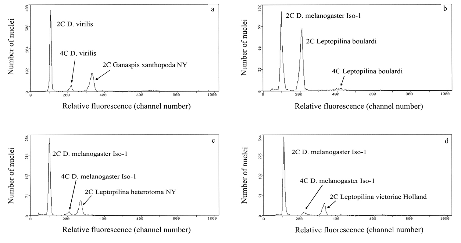
Cytograms showing relative fluorescence and total propidium iodide-stained nuclei of samples and standards to determine genome size. a relative fluorescence of PI-stained 2C nuclei from one head of a Ganaspis xanthopoda strain NY female co-prepared with 2C and 4C nuclei from one head of a Drosophila virilis female standard (1C = 333 Mb) b–d relative fluorescence and total PI stained nuclei of co-prepared Leptopilina and Drosophila melanogaster (1C = 175 Mb) to determine genome size for Leptopilina boulardi (panel b), Leptopilina heterotoma (panel c), and Leptopilina victoriae (panel d). Genome size is calculated as follows: (mean fluorescence channel number of sample 2C peak/mean fluorescence channel number of 2C standard peak) X 1C DNA content of the standard, with the genome size mean and standard error calculated from repeat co-preparations using different individuals of each species.

Our results provide the first information on genome sizes not only of the family Figitidae, but of the superfamiy Cynipoidea as a whole. It is intriguing that the genome sizes of all these parasitoids exceed the average value known for previously studied Hymenoptera, i.e., 360.75 Mb (Tsutsui et al. 2008, Ardila-Garcia et al. 2010), but are fairly close to those of many Chalcidoidea (Tsutsui et al. 2008, Ardila-Garcia et al. 2010), the closest group to cynipoids (see Sharkey 2007).

Karyotypes

Total lengths of haploid chromosome sets of above mentioned species are given in Table 1. The relative lengths and centromere indices of all chromosomes are given in Table 2.

Table 2.

Relative lengths (RL) and centromere indices (CI) of Drosophila parasitoids. (mean±SE). Strains and numbers of studied metaphase plates are as in Table 1. Centromere indices are: metacentrics: 37.5-50.0; submetacentrics: 25.0-37.5; subtelocentrics: 12.5-25.0; acrocentrics: 0-12.5, according to Levan et al. (1964).

Species/ chromosome no. Ganaspis xanthopoda Leptopilina boulardi Leptopilina heterotoma Leptopilina victoriae
RL CI RL CI RL CI RL CI
1 24.17± 0.77 37.50± 5.34 31.13± 0.81 39.97± 2.98 14.21± 0.32 28.26± 1.42 15.49± 0.21 39.27± 4.55
2 12.85± 0.25 20.50± 1.57 13.06± 0.37 35.13± 2.79 11.89± 0.16 30.03± 1.07 11.63± 0.20 30.46± 4.97
3 11.97± 0.11 20.14± 1.28 11.45± 0.27 29.86± 2.56 11.01± 0.11 28.58± 1.44 11.04± 0.17 32.95± 5.05
4 10.59± 0.32 19.80± 3.50 9.19± 0.17 21.03± 2.99 10.51± 0.82 27.72± 1.16 10.48± 0.19 31.88± 4.34
5 9.35± 0.61 22.25± 3.56 8.54± 0.19 18.66± 4.06 10.02± 0.69 28.90± 1.93 9.53± 0.13 33.69± 5.72
6 8.75± 0.43 35.55± 2.49 7.32± 0.19 17.33± 2.79 9.40± 0.11 33.17± 1.89 9.15± 0.13 31.12± 3.84
7 8.39± 0.15 15.86± 0.39 6.95± 0.15 11.86± 3.68 8.92± 0.11 32.12± 1.96 9.02± 0.11 34.72± 4.95
8 7.81± 0.42 43.48± 1.32 6.42± 0.10 13.91± 4.84 8.48± 0.12 30.16± 1.54 8.69± 0.08 41.12± 2.45
9 6.12± 0.10 1.44± 0.73 5.94± 0.13 8.77± 2.60 8.04± 0.11 28.70± 1.49 8.01± 0.26 34.88± 2.57
10 - - - - 7.52± 0.09 31.93± 1.88 6.96± 0.28 36.19± 1.87

Ganaspis xanthopoda. Nine chromosomes were found in the haploid karyotype of this species (n = 9; Fig. 2a). Chromosomes are long relative to Leptopilina spp. (see Table 1 and below); most of them are of similar size. However, the first meta- or submetacentric chromosome is about twice as long as the remaining ones. Most other chromosomes are subtelocentric, except for the sixth submetacentric, eighth metacentric, and last acrocentric ones.

Figure 2.

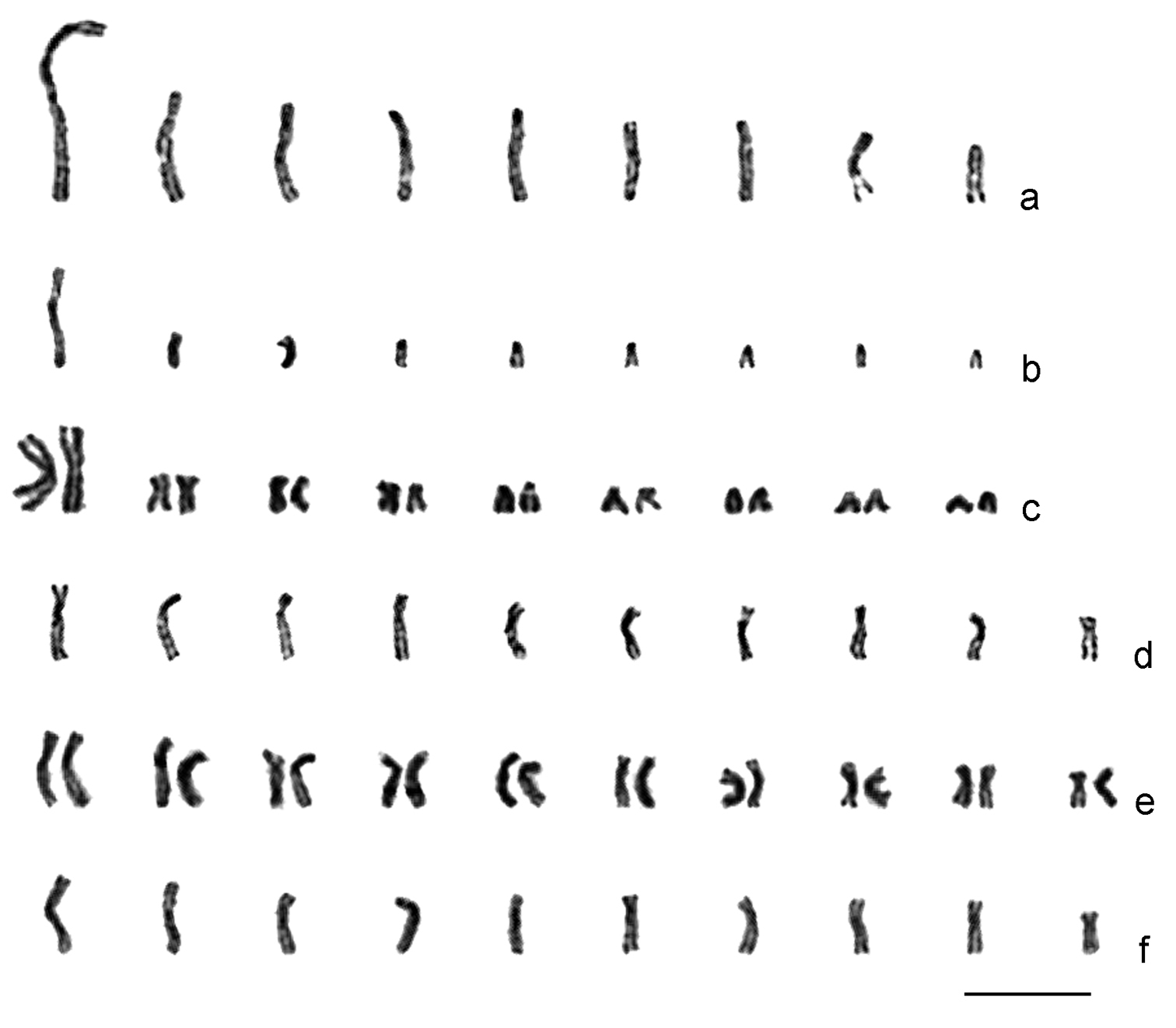
Karyograms of Drosophila parasitoids. a Ganaspis xanthopoda, haploid set b Leptopilina boulardi (strain 17), haploid set c ditto, diploid set d Leptopilina heterotoma (New York strain), haploid set e ditto, diploid set f Leptopilina victoriae, haploid set. Scale bar 10 μm.

Leptopilina boulardi. As in Ganaspis xanthopoda, n = 9 (and 2n = 18; Figs 2b and c; Fig. 3a). Moreover, the karyotype of Ganaspis xanthopoda is superficially similar to that of Leptopilina boulardi in that the very large first metacentric chromosome is more than twice as long as the second. However, the length of all remaining Leptopilina boulardi chromosomes is roughly half that of the Ganaspis xanthopoda chromosomes. Furthermore, chromosomes of the second and third pairs are submetacentric, those of the fourth, fifth, sixth and eighth pairs are subtelocentric, and chromosomes of the seventh and ninth pairs are acrocentric.

Figure 3.

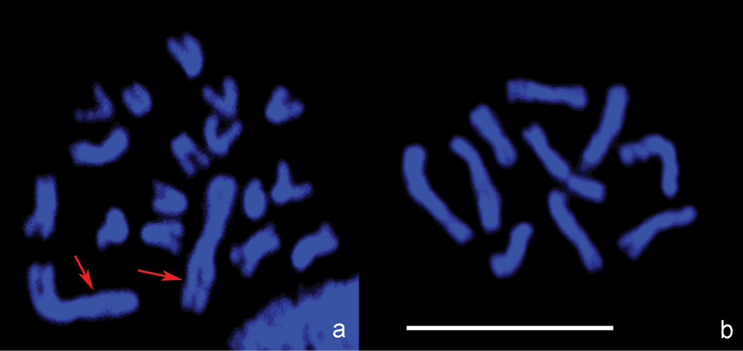
Confocal microscopic images. a Leptopilina boulardi, diploid metaphase plate b- Leptopilina heterotoma, haploid metaphase plate. Arrows point to the pair of large metacentric chromosomes in the karyotype of Leptopilina boulardi that presumably arose via chromosomal fusion in an ancestral chromosome set with n = 10. Scale bar 10 μm.

Leptopilina heterotoma. Consistent with previous observations (Crozier 1975), we found n = 10 and 2n = 20 in this species (Figs 2d and e; Fig. 3b). All chromosomes of the karyotype are submetacentrics that gradually decrease in size.

Leptopilina victoriae. This species belongs to the Leptopilina heterotoma clade (Allemand et al. 2002), and unsurprisingly, its karyotype is similar to that of Leptopilina heterotoma. The haploid karyotype of Leptopilina victoriae contains ten submetacentric or metacentric chromosomes (n = 10) of similar size (Fig. 2f). The first chromosome of Leptopilina victoriae is significantly longer and the fifth and tenth chromosomes are significantly shorter than the corresponding chromosomes of Leptopilina heterotoma. In addition, the centromere position in the first and eighth submetacentric chromosomes is significantly different than observed for the apparently metacentric chromosomes of Leptopilina victoriae.

Discussion

Parasitic wasps make up a significant number of species of all insects (LaSalle and Gauld 1993). However, because of the complete absence of genomic information, the molecular biology and genetics of parasitic wasps of Drosophila have lagged behind, even though it is now possible to rapidly sequence genomes of organisms without prior genetic or genomic information.

In a study of genome size of 89 species of bees, wasps, and ants, Ardila-Garcia et al. (2010) hypothesized that genome sizes are constrained by traits associated with parasitism or eusociality. They however found that not all parasitoids have small genomes (Ardila-Garcia et al. 2010). So while it is not altogether surprising that the genomes of the koinobiont parasitoids of Drosophila studied here are as large as that of nonparasitic Hymenoptera, it is intriguing that they have such large genomes. Koinobionts keep their host alive; and must develop and emerge before their host is exhausted and dies. Small genomes replicate faster and require fewer resources, which imposes a selection cost on a bloated genome. An antagonist selective force must act on the parasitoid genome. Because of their obligate and intimate relationship with their fly hosts, it is possible that parasitic wasps take up, or share genetic information via transposons. Widespread transfer of genes laterally has recently been documented from Wolbachia Hertig, 1936 to insect or nematode genomes (Hotopp et al. 2007). Wolbachia has been associated with many parasitic wasps of Drosophila (Vavre et al. 2009). Genomic sequence information will reveal if horizontal transmission of transposons, facilitated by the parasitic life style, may have contributed to the large genome size. In this scenario, different transposon types, with rapid turnover in the genome are expected.

Our karyotypic study provides new insights into the genome structure of Drosophila parasitoids. First, the study demonstrates an obvious positive correlation between the genome size and total chromosome length in those parasitic wasps (Table 1; Fig. 4). However, chromosome length in Ganaspis xanthopoda increases relatively slower than might be expected from its larger genome size (Fig. 4). This observation suggests that a significant portion of the bloated Ganaspis genome is repeat sequence that is highly condensed at metaphase. High copy number of satellite DNA is associated with genome size variation in Drosophila species (Bosco et al. 2007) and it is possible that a similar discrepancy in transposon or satellite DNA in the Ganaspis xanthopoda genome accounts for smaller than expected increase in chromosome length (Fig. 4).

Figure 4.

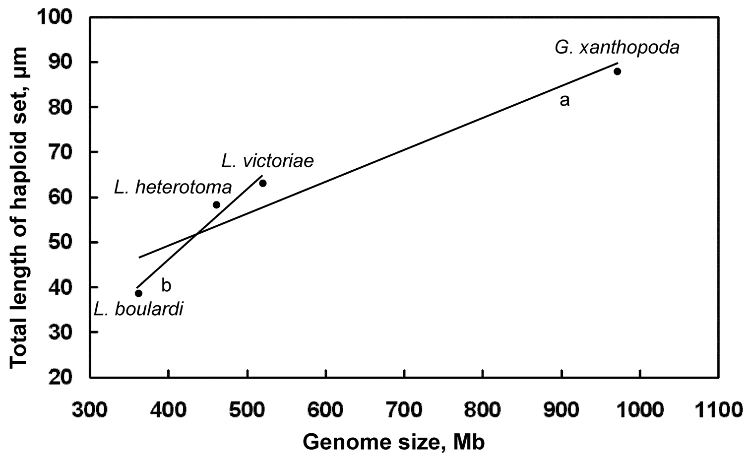
Distribution of genome size/chromosome length of Drosophila parasitoids. Mean values are given for each species. Trend lines: a for all species combined b for Leptopilina spp. (i.e. all species excluding Ganaspis xanthopoda).

Second, our study reveals that genome sizes vary independently of the chromosome number in Drosophila parasitoids. This may not be surprising if the large metacentric chromosomes of Leptopilina boulardi (Fig. 2b; Fig. 3a) and Ganaspis xanthopoda (Fig. 2a) have an independent origin via chromosomal fusions. Parallel chromosomal fusions are relatively frequent within various lineages of parasitic Hymenoptera (Gokhman 2004, 2009). In addition to Ganaspis and Leptopilina, chromosome numbers of Phaenoglyphis villosa (Hartig, 1841) (n = 10) and Callaspidia defonscolombei Dahlbom, 1842 (n = 11) from the same family have been studied (see Gokhman 2009). This information indicates that n = 10 (or a value close to 10) is likely to be an initial chromosome number for species of the Leptopilina/Ganaspis clade. If this is true, karyotypes with n = 9 found in Ganaspis xanthopoda and Leptopilina boulardi as well as that with n = 5 found in Leptopilina clavipes are likely to have resulted from chromosomal fusions and are therefore derived from a chromosome set that was probably similar to the karyotypes of Leptopilina heterotoma or Leptopilina victoriae (see also e.g. Gokhman 2010).

Third, the karyotype provides the scaffold number for future sequencing effort in these insects. When the karyotypic features of the species studied here are superimposed onto their phylogeny (Schilthuizen et al. 1998), clear correspondence is revealed: Leptopilina heterotoma and Leptopilina victoriae share very similar karyotypes, and are the most closely related species. In contrast, Leptopilina boulardi belongs to a distinct clade of the Leptopilina genus. Cytogenetic mapping of Expressed Sequence Tags, combined with restriction-site associated DNA (RAD) sequencing (Baird et al. 2008) based on the karyotype would ensure the highest quality genomic assembly, and pave the way for comparative genomics of parasitoid wasps of Drosophila. Such comparative genomics will provide insights into the organization of the host and parasitoid genomes and the co-evolution of these insects in nature.

Acknowledgements

The authors are grateful to Andrey P. Mikhailenko (MSU Botanical Garden) for technical assistance. This work was supported by grants from the Russian Foundation for Basic Research (10-04-01521) to V.E.G., and from the U.S. Department of Agriculture (NRI/USDA CSREES 2006-03817 and NIFA 2009-35302-05277); National Institute of Health (NIGMS S06 GM08168, GM056833-10, P50-GM68762, and G12-RR03060); and the Professional Staff Congress of the City University of New York (S.G.), and a National Human Genome Research Institute (NHGRI) subcontract to J.S.J. The authors declare no conflict of interest.

References
Allemand R, Lemaître C, Frey F, Boulétreau M, Vavre F, Nordlander G, van Alphen J, Carton Y (2002) Phylogeny of six African Leptopilina species (Hymenoptera: Cynipoidea, Figitidae), parasitoids of Drosophila, with description of three new species. Annales de la Société entomologique de France 38: 319-332.
Ardila-Garcia AM, Umphrey GJ, Gregory TR (2010) An expansion of the genome size dataset for the insect order Hymenoptera, with a first test of parasitism and eusociality as possible constraints. Insect Molecular Biology 19: 337–346. doi: 10.1111/j.1365-2583.2010.00992.x.
Baird NA, Etter PD, Atwood TS, Currey MC, Shiver AL, Lewis ZA, Selker EU, Cresko WA, Johnson EA (2008) Rapid SNP discovery and genetic mapping using sequenced RAD markers. PLoS One 3: e3376. doi: 10.1371/journal.pone.0003376
Bosco G, Campbell P, Leiva-Neto JT, Markow TA (2007) Analysis of Drosophila species genome size and satellite DNA content reveals significant differences among strains as well as between species. Genetics 177: 1277–1290. doi: 10.1534/genetics.107.075069
Chiu H, Morales J, Govind S (2006) Identification and immuno-electron microscopy localization of p40, a protein component of immunosuppressive virus-like particles from Leptopilina heterotoma, a virulent parasitoid wasp of Drosophila. Journal of General Virology 87: 461–470. doi: 10.1099/vir.0.81474-0
Crosby MA, Goodman JL, Strelets VB, Zhang P, Gelbart WM (2007) FlyBase: genomes by the dozen. Nucleic Acids Research 35 (supplement 1): D486-D491. doi: 10.1093/nar/gkl827
Crozier RH (1975) Hymenoptera. Animal Cytogenetics. Gebrüder Borntraeger, Berlin-Stuttgart, 95 pp.
Flybase. A Database of Drosophila Genes & Genomes, FB2011_04, released April 22nd, 2011. http://flybase.org/ [accessed 29 April 2011].
Gokhman VE (2004) Karyotype evolution in parasitic Hymenoptera. Entomological Review 84 (supplement): S41-S49.
Gokhman VE (2009) Karyotypes of Parasitic Hymenoptera. Springer Science+Business Media B.V., Dordrecht, 183 pp. doi: 10.1007/978-1-4020-9807-9
Gokhman VE (2010) Chromosomes of parasitic wasps of the genus Metaphycus (Hymenoptera: Chalcidoidea: Encyrtidae). Comparative Cytogenetics 4: 21–25. doi: 10.3897/compcytogen.v4i1.29
Gregory TR, Johnston JS (2008) Genome size diversity in the family Drosophilidae. Heredity 101: 228–238. doi: 10.1038/hdy.2008.49
Hotopp JCD, Clark ME, Oliveira DCSG, Foster JM, Fisher P, Torres MCM, Giebel JD, Kumar N, Ishmael N, Wang S, Ingram J, Nene RV, Shepard J, Tomkins J, Richards S, Spiro DJ, Ghedin E, Slatko BE, Tettelin H, Werren JH (2007) Widespread lateral gene transfer from intracellular bacteria to multicellular eukaryotes. Science 317: 1753–1756. doi: 10.1126/science.1142490
Imai HT, Taylor RW, Crosland MWJ, Crozier RH (1988) Modes of spontaneous chromosomal mutation and karyotype evolution in ants with reference to the minimum interaction hypothesis. Japanese Journal of Genetics 63: 159-185.
Johnston JS, Ross LD, Beani L, Hughes DP, Kathirithamby J (2004) Tiny genomes and endoreduplication in Strepsiptera. Insect Molecular Biology 13: 581–585. doi: 10.1111/j.0962-1075.2004.00514.x
Kraaijeveld AR, Godfray HCJ (2009) Evolution of host resistance and parasitoid counter-resistance. Advances in Parasitology 70: 257–280. doi: 10.1016/s0065-308x(09)70010-7
LaSalle J, Gauld ID (1993) Hymenoptera: their biodiversity, and their impact on the diversity of other organisms. In: LaSalle J, Gauld ID (Eds). Hymenoptera and Biodiversity. CAB International, Wallingford: 1-26.
Lanot R, Zachary D, Holder F, Meister M (2001) Postembryonic hematopoiesis in Drosophila. Developmental Biology 230: 243–257. doi: 10.1006/dbio.2000.0123
Lee MJ, Kalamarz ME, Paddibhatla I, Small C, Rajwani R, Govind S (2009) Virulence factors and strategies of Leptopilina spp.: selective responses in Drosophila hosts. Advances in Parasitology 70: 123–145. doi: 10.1016/s0065-308x(09)70005-3
Levan A, Fredga K, Sandberg AA (1964) Nomenclature for centromeric position on chromosomes. Hereditas 52: 201-220.
Melk JP, Govind S (1999) Developmental analysis of Ganaspis xanthopoda, a larval parasitoid of Drosophila melanogaster. Journal of Experimental Biology 202: 1885-1896.
Paddibhatla I, Lee MJ, Kalamarz ME, Ferrarese R, Govind S (2010) Role for sumoylation in systemic inflammation and immune homeostasis in Drosophila larvae. PLoS Pathogens 6: e1001234. doi: 10.1371/journal.ppat.1001234
Pannebakker BA, Pijnacker LP, Zwaan BJ, Beukeboom LW (2004) Cytology of Wolbachia-induced parthenogenesis in Leptopilina clavipes (Hymenoptera: Figitidae). Genome 47: 299–303. doi: 10.1139/g03-137
Schilthuizen M, Nordlander G, Stouthamer R, van Alphen J (1998) Morphological and molecular phylogenetics in the genus Leptopilina (Hymenoptera: Cynipoidea: Eucoilidae). Systematic Entomology 23: 253–264. doi: 10.1046/j.1365-3113.1998.00049.x
Schlenke TA, Morales J, Govind S, Clark AG (2007) Contrasting infection strategies in generalist and specialist wasp parasitoids of Drosophila melanogaster. PLoS Pathogens 3: e158. doi: 10.1371/journal.ppat.0030158
Sharkey M (2007) Phylogeny and classification of Hymenoptera. Zootaxa 1668: 521-548.
Sorrentino RP, Melk JP, Govind S (2004) Genetic analysis of contributions of dorsal group and JAK-Stat92E pathway genes to larval hemocyte concentration and the egg encapsulation response in Drosophila. Genetics 166: 1343–1356. doi: 10.1534/genetics.166.3.1343
StatSoft Inc (1995) STATISTICA for Windows. Computer Program Manual.
Tsutsui ND, Suarez AV, Spagna JC, Johnston JS (2008) The evolution of genome size in ants. BMC Evolutionary Biology 8: 64. doi: 10.1186/1471-2148-8-64
Vavre F, Mouton L, Pannebakker BA (2009) Drosophila-parasitoid communities as model systems for host-Wolbachia interactions. Advances in Parasitology 70: 299–331. doi: 10.1016/s0065-308x(09)70012-0