|
||
|
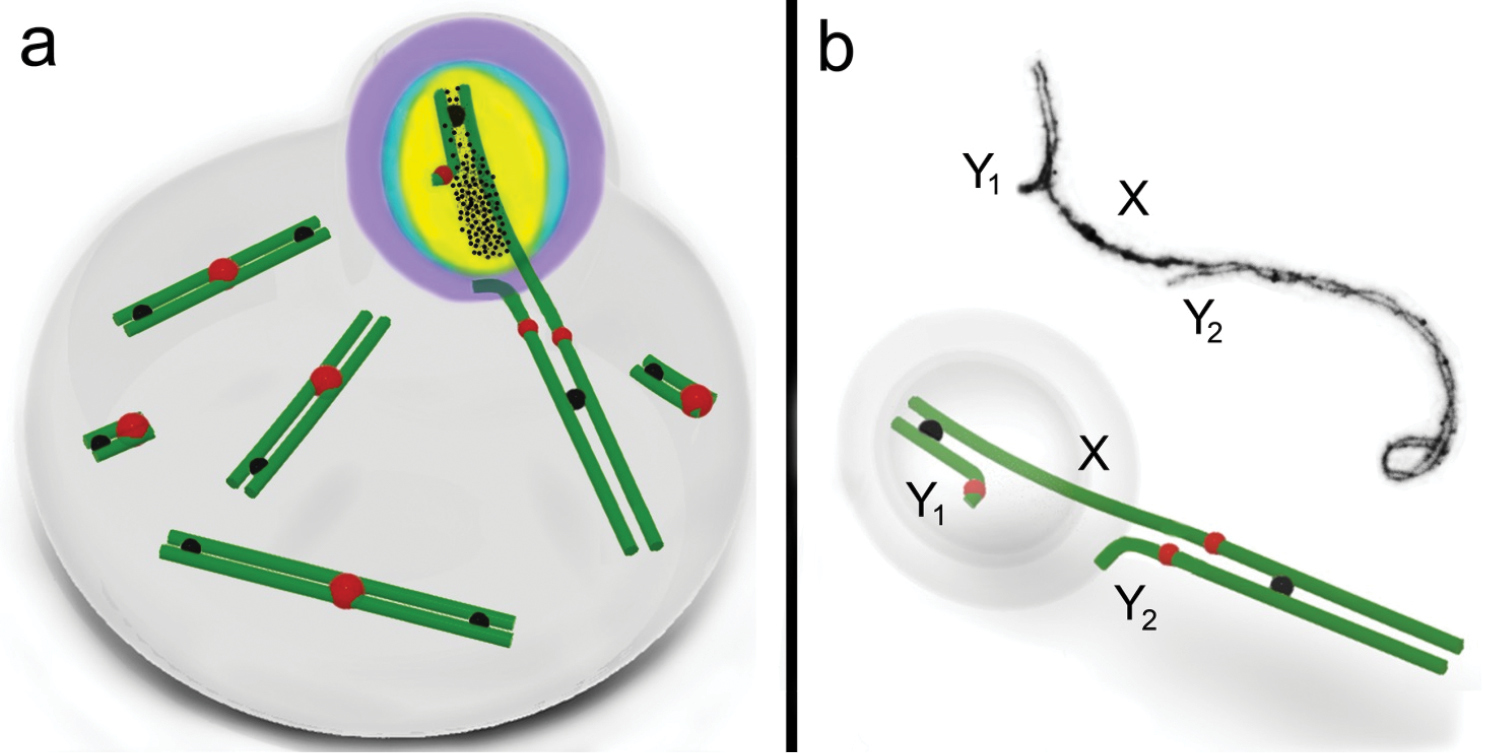
Schematic illustration of male common shrew MSCI. A mid-pachytene spermatocyte (a) and a sex (XY1Y2) trivalent (b) of a shrew are shown. An electron micrograph of the sex trivalent is shown at the top of the b. The true sex chromosome regions (part of the X and the Y1) form a sex body on the periphery of the nucleus. The chromatin of the sex body undergoes reorganisation. MSCI markers have different distributions: SUMO-1 (yellow), ATR (black dots), ubiH2A (blue), γH2AFX (violet). ATR is localised on the true sex chromosome regions, and is especially intense on the asynaptic region with a smaller amount where there is synapsis. SUMO-1 and ubiH2A are localised on both the asynaptic and synaptic regions of the true sex chromosome regions. γH2AFX overlays all the true sex chromosome regions and the unpaired part of the Y2 axial element. Representative autosomal SCs are shown. MLH1 signals are shown as black balls. The red balls indicate centromeres. |