|
||
|
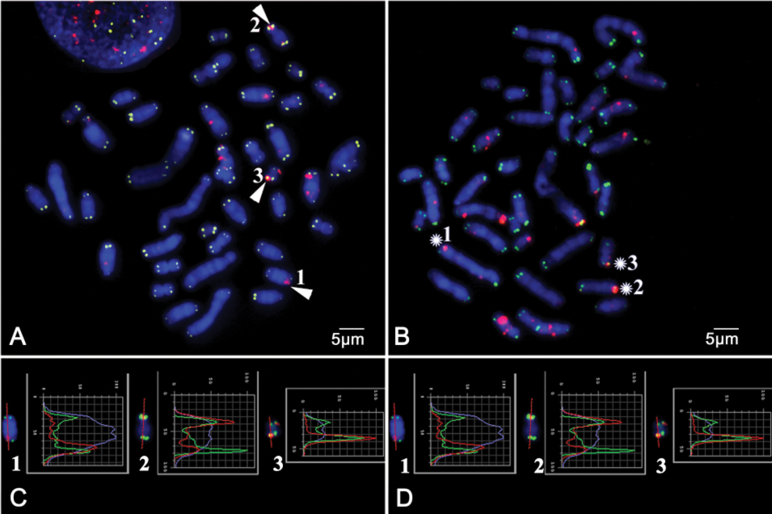
Immunochemistry with antibodies to ɤ-H2AX (red) and further FISH with telomeric PNA probe (CCCTAA)3 conjugated with FITC (green). Chromosomes stained DAPI (blue) A The metaphase spread of RNFF1 cell line on 9th passage B The metaphase spread of MR39 cell line on 6th passage. The arrows and asterisks indicate dysfunctional telomeres C – D. Curves of signal intensities along the length of telomeres containing dysfunctional telomeres on individual chromosomes from A–B respectively. Scale bars: 5 µm |