Comparative Cytogenetics 5(4): 329-344, doi: 10.3897/CompCytogen.v5i4.1149
Heterochromatin heterogeneity in Hypostomus prope unae (Steindachner, 1878)(Siluriformes, Loricariidae)from Northeastern Brazil
J.A. Bitencourt 1, P.R.A.M. Affonso 2, L. Giuliano-Caetano 1, A.L. Dias 1
1 Departamento de Biologia Geral, Universidade Estadual de Londrina, CCB, Londrina - 86051-970, Paraná, Brazil
2 Departamento de Ciências Biológicas, Universidade Estadual do Sudoeste da Bahia, DCB, Jequié - 45200-000, Bahia, Brazil

Corresponding author: P.R.A.M. Affonso (paulomelloaffonso@yahoo.com.br)

Academic editor: S. Grozeva

received 17 February 2011 | accepted 21 April 2011 | Published 9 November 2011


(C) 2011 J.A. Bitencourt. This is an open access article distributed under the terms of the Creative Commons Attribution License, which permits unrestricted use, distribution, and reproduction in any medium, provided the original author and source are credited.


For reference, use of the paginated PDF or printed version of this article is recommended.

Abstract

Cytogenetic analyses using C-banding and chromosomal digestion by several restriction enzymes were carried out in four populations (named A, B, C and D) of Hypostomus prope unae (Loricariidae, Hypostominae) from Contas river basin, northeastern Brazil. These populations share 2n=76 and single NORs on the second metacentric pair but exclusive karyotype forms for each locality. Populations A and B presented conspicuous terminal and interstitial heterochromatic blocks on most of acrocentric chromosomes and equivalent to NORs with differences in both position and bearing pair. Population D showed evident marks at interstitial regions and interspersed with nucleolar region while population C presented interstitial and terminal heterochromatin segments, non-coincident with NORs. The banding pattern after digestion with the endonucleases Alu I, Bam HI, Hae III and Dde I revealed a remarkable heterogeneity within heterochromatin, allowing the identification of distinctive clusters of repeated DNA in the studied populations, besides specific patterns along euchromatic regions. The analysis using restriction enzymes has proved to be highly informative, characterizing population differences and peculiarities in the genome organization of Hypostomus prope unae.

Keywords

C-banding, heterochromatin, ichthyofauna, restriction enzymes

Introduction

Restriction enzymes (RE) represent a powerful tool for studies about DNA organization (Lima-de-Faria et al. 1980). Such bacterial endonucleases recognize and cleavage target-sequences in the double-strand DNA, providing a highly specific pattern of chromosomal banding according to each enzyme (Lloyd and Thorgaard 1988). The removal of DNA fragments allows studying both structure and base composition of specific chromosomal regions (Lorite et al. 1999; Sánchez et al. 1990, 1991; Lozano et al. 1991; Bianchi et al. 1985). Therefore, the RE banding pattern is an exceptionally sensitive method in heterochromatin analysis (Pieczarka et al. 1996), being able to reveal a higher degree of heterogeneity and more refine comparative analyses than the traditional C-banding itself.

In spite of the intensive application of restriction enzymes in chromosomal analyses of several animal groups (Miller et al. 1976; Kaelbling et al. 1984; Lima-de-Faria et al. 1980; Bianchi et al. 1985; Marchi and Mezzanotte 1988, 1990; Juan et al. 1990; Pieczarka et al. 1996), a few studies of RE-based heterochromatin differentiation are reported in fish chromosomes, being restricted to some groups such as Characidae (Kantek et al. 2007; Maistro et al. 1999), Prochilodontidae (Maistro et al. 2000), Pimelodidae (Swarça et al. 2005; Carvalho and Dias 2005), Salmonidae (Lloyd and Thorgaard 1988; Sánchez et al. 1990, 1991; Lozano et al. 1991; Albuín et al. 1994), Muraenidae (Cau et al. 1988) and Scophthalmidae (Bouza et al. 1994).

Within the genus Hypostomus Lacépède, 1803, heterochromatin can be associated to heteromorphic chromosomes (Cereali et al. 2008; Kavalco et al. 2004, 2005), sex chromosomes (Artoni et al. 1998) and polymorphism cases (Rubert et al. 2008). In addition, species of this genus usually present a remarkable variability in both distribution and composition of heterochromatin (Artoni and Bertollo 1999). However, these data refer to C-banding and/or fluorochrome staining while studies using enzymatic digestion have not been reported in the genus or the family Loricariidae so far.

The goal of the present work was to analyze comparatively metaphase chromosomes of Hypostomus prope unae (Steindachner, 1878) by C-banding and RE digestion in order to refine previous cytogenetic studies (Bitencourt 2010) among four populations of this species along a poorly studied coastal river basin in northeastern Brazil.

Methods

Forty-six specimens of Hypostomus prope unae from four collection sites in Contas river basin were analyzed, being 10 (3 males, 2 females and 5 immature) from the main channel of Contas river (13°51'51"S, 40°04'54"W), 10 (6 males, 1 female and 3 immature) from Preto do Costa river (13°45'84"S, 39°56'47"W), 15 (9 males and 6 immature) from Oricó river (14°08'03"S, 39°21'30"W), and 11 (4 males, 4 females and 3 immature) from Preto do Criciúma river (13°55'45"S, 39°57'57"W) (Fig. 1).

Figure 1.

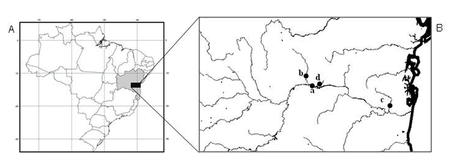
A–B Collection sites A Map of Brazil, highlighting the state of Bahia in northeastern region; B Contas river basin and respective sampling sites: a- Contas river, b- Preto do Costa river, c- Oricó river, d- Preto do Criciúma river.

Voucher specimens were identified by Dr. Claudio Zawadski from Universidade Estadual de Maringá (UEM) and deposited in the fish collection at NUPELIA – UEM, Maringá, PR, Brazil (NUP 9811, 9814). These four populations are referred as A, B, C and D, respectively.

Metaphase chromosomes were obtained from kidney cells as described by Bertollo et al. (1978) after mitotic stimulation using yeast suspension (Lee and Elder 1980) or, alternatively, Munolan® (bacterial and fungal antigens) diluted in water (1 pill per 0.5mL of water), as suggested by Molina (2001). The chromosomes were classified into metacentric (m), submetacentric (sm), subtelocentric (st) and acrocentric (a), as commonly described in fish (Levan et al. 1964). The fundamental number (FN) was established taking into account that m, sm and st chromosomes are bi-armed while chromosomes bear one chromosomal arm.

C-positive heterochromatin was detected according to Sumner (1972), with slight modifications. In situ digestion using restriction enzymes was performed as proposed by Mezzanotte et al. (1983), with modifications. Concentration and incubation tests were extensively performed to optimize the results. After defining the best concentration (Table 1), we added 30 μl of each enzyme solution (diluted in specific buffer and distilled water) onto chromosomal preparations. The slides were incubated in moist chamber at 37°C for specific periods according to each enzyme (Table 1). Afterwards, the slides were washed in distilled water and stained with 5% Giemsa in phosphate buffer (pH 6.8) for 8 minutes.

Table 1.

List of restriction endonucleases (RE) used on the chromosomal preparations of Hypostomus prope unae, with their respective restriction sites and optimum concentrations and incubation periods obtained in the present work.

Endonucleases Restriction site Concentration Incubation
Alu I (5’- AG CT - 3’) 0.4 U/ µl 4h
Bam HI (5’- G GATCC - 3’) 0.5 U/ µl 15h
Hae III (5’-GG CC - 3’) 0.6 U/ µl 14h
Dde I (5’- C TNAG – 3’) 2 U/ µl 4h
Results

The specimens from all analyzed populations presented a modal chromosomal number of 2n=76 and distinct karyotype formulae, as follows: 12m+16sm+48st/a (FN= 104) for specimens from population A, 12m+20sm+44st/a (FN=108) for specimens from population B, 10m+14sm+52st/a (FN=100) for individuals from population C and 10m+20sm+46st/a (FN= 106) for those from population D. Furthermore, distinctive patterns of heterochromatin distribution were detected by C-banding. Although populations A and B bear conspicuous terminal and interstitial marks in 17 chromosomal pairs as well as centromeric and NOR-associated heterochromatin, they differ in relation to C-bands position or bearing pair (Figs 2, 3).

Figure 2.

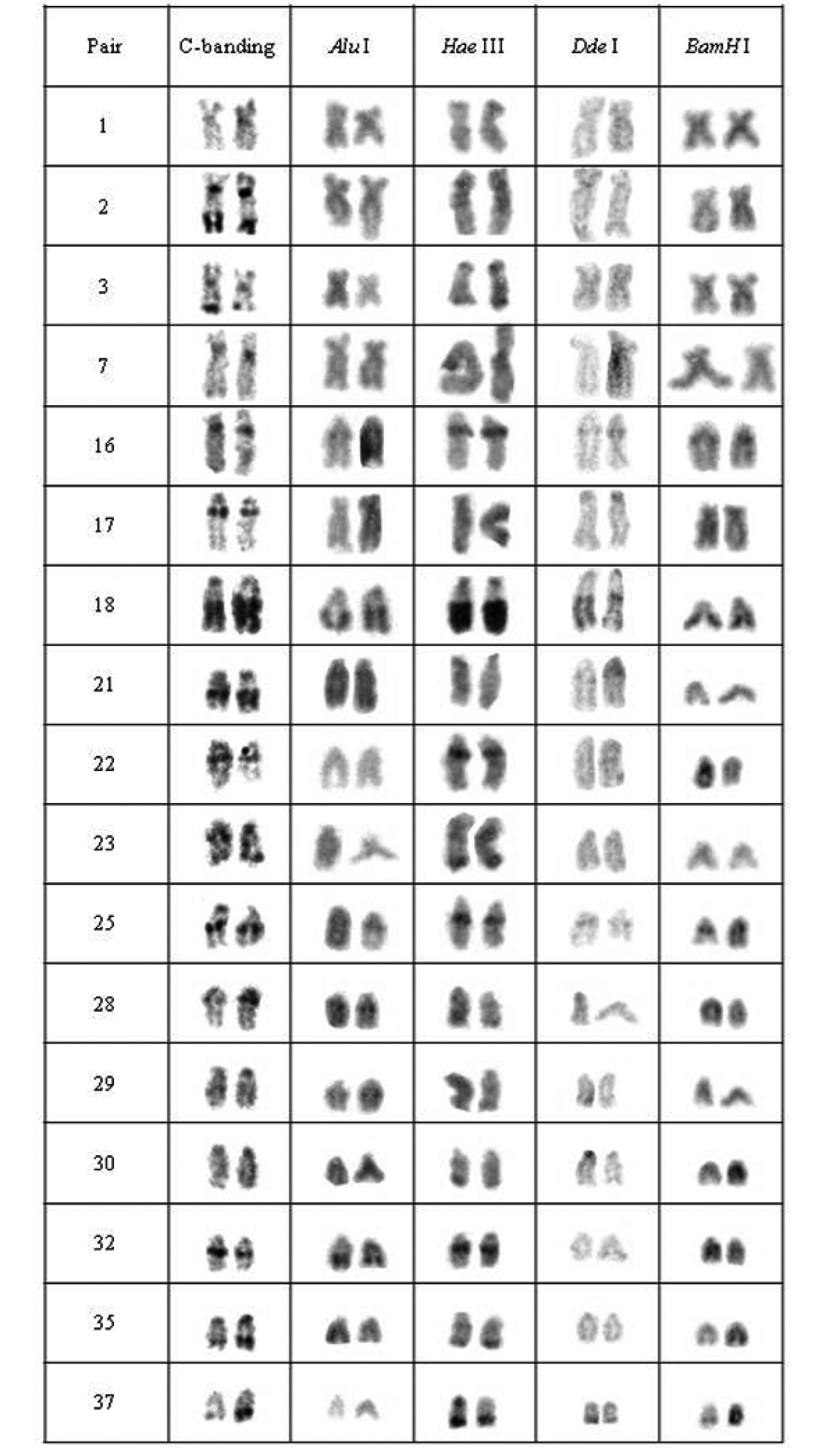
Chromosomal pairs from population A of Hypostomus prope unae showing the C-positive heterochromatin and banding pattern after digestion with the restriction endonucleases: Alu I, Hae III, Dde I and Bam HI.

Figure 3.

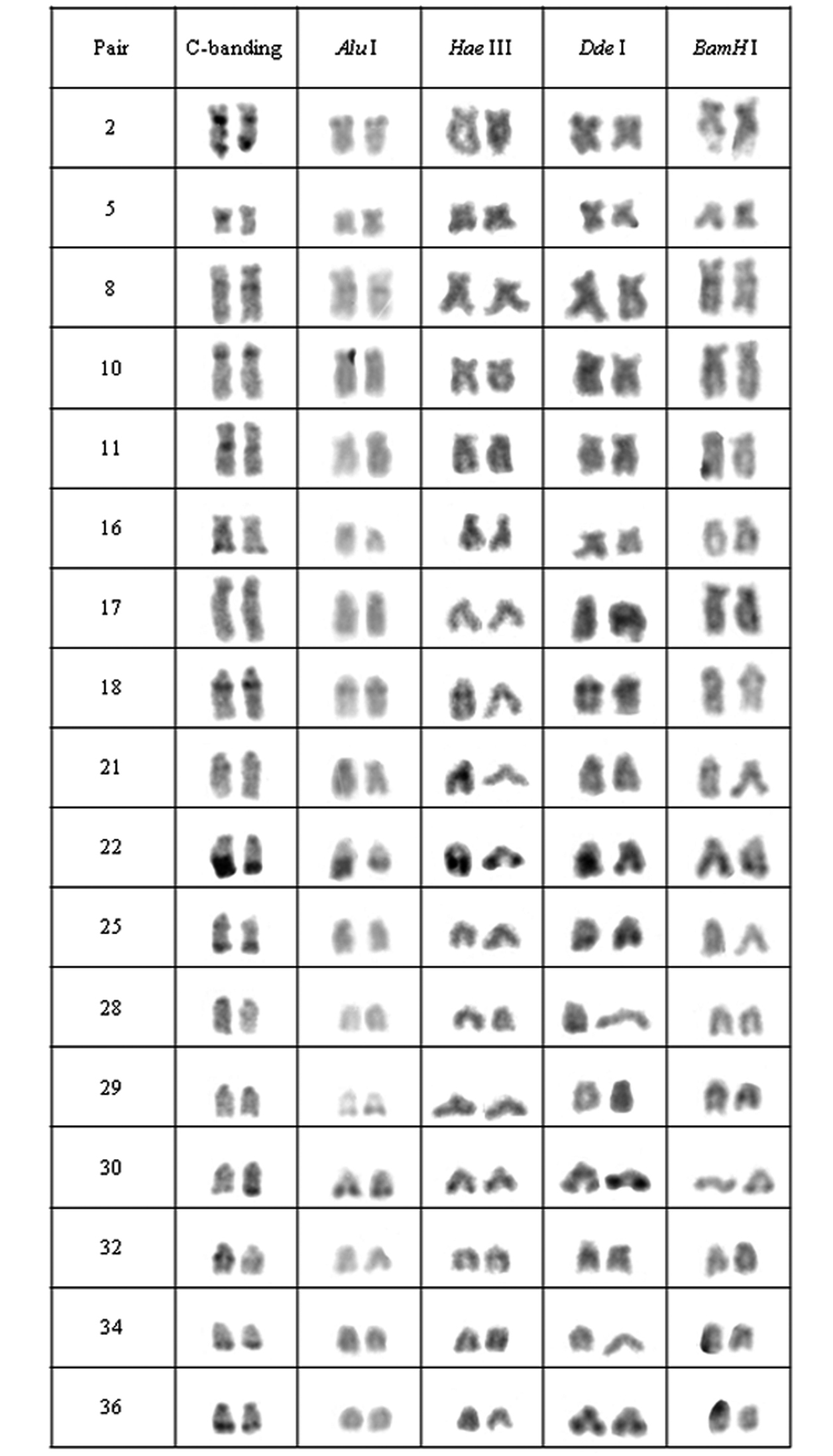
Chromosomal pairs from population B of Hypostomus prope unae showing the C-positive heterochromatin and banding pattern after digestion with the restriction endonucleases: Alu I, Hae III, Dde I and Bam HI.

Heteromorphic blocks were also evident in both populations. Besides the NOR-bearing pair, 18th, 21st and 37th pairs in population A and the 22nd pair in population B size differences between homologous (Figs 2, 3). Population C was characterized by interstitial and terminal marks in six chromosomal pairs, non-coincident with NORs (Fig. 4). On the other hand, population D presented eight pairs, most of them acrocentric, bearing interstitial C-bands and also interspersed with NORs (Fig. 5).

Figure 4.

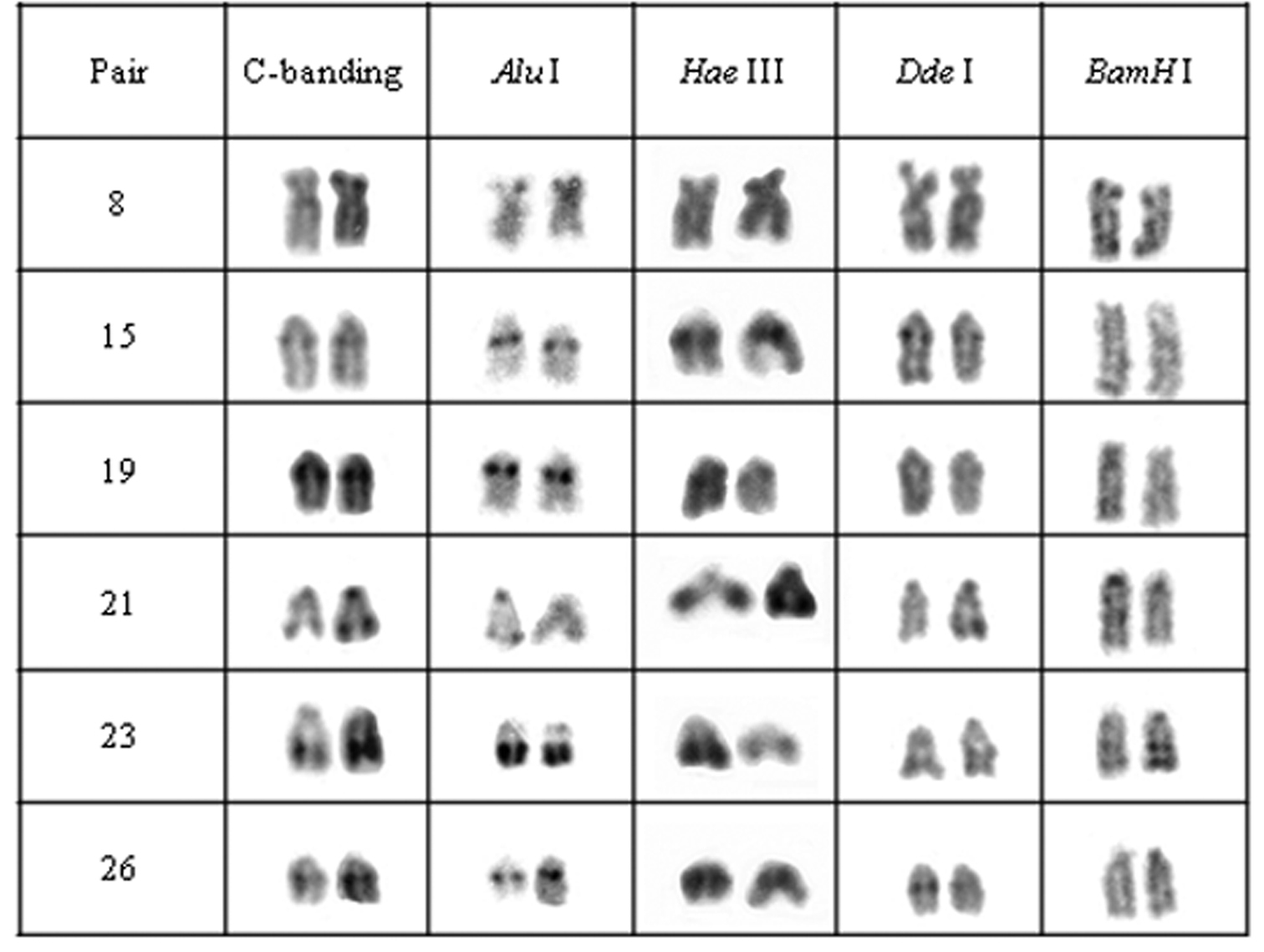
Chromosomal pairs from population C of Hypostomus prope unae showing the C-positive heterochromatin and banding pattern after digestion with the restriction endonucleases: Alu I, Hae III, Dde I and Bam HI.

Figure 5.

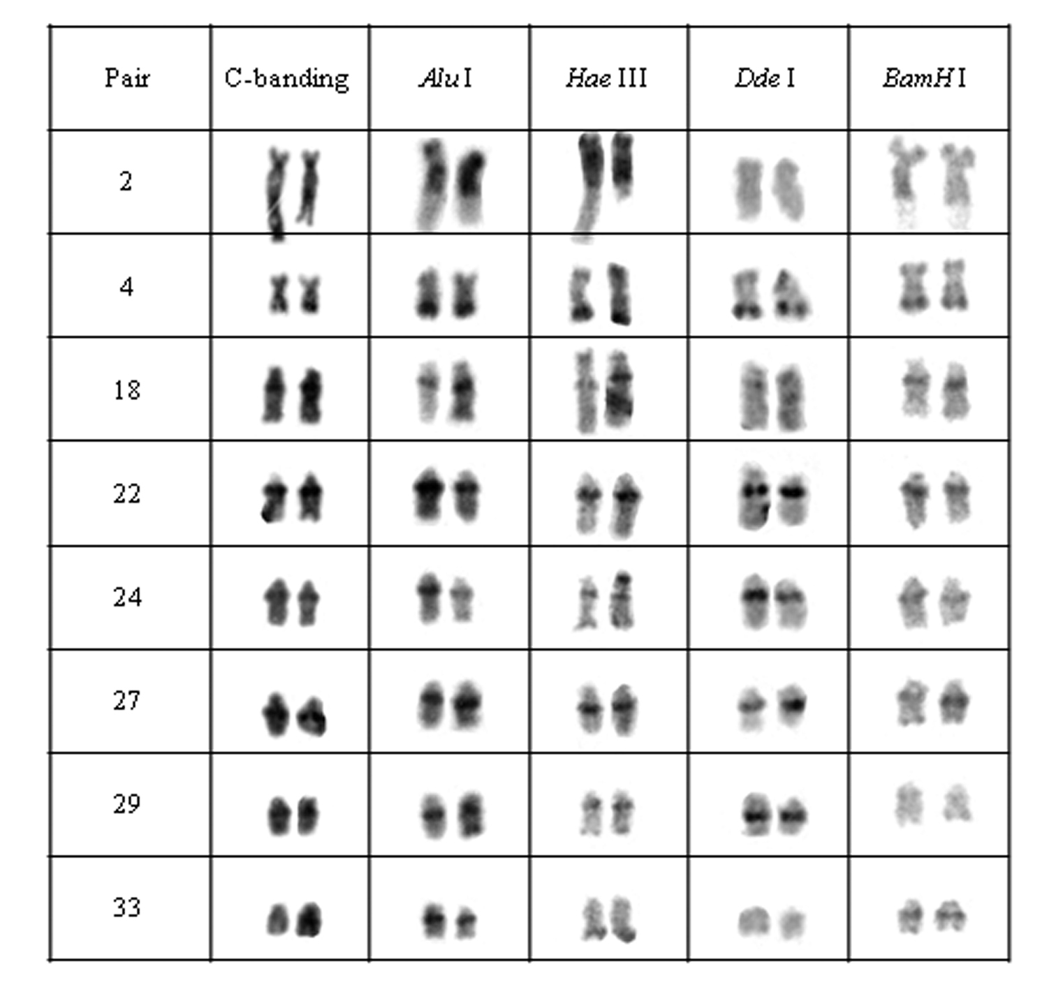
Chromosomal pairs from population D of Hypostomus prope unae showing the C-positive heterochromatin and banding pattern after digestion with the restriction endonucleases: Alu I, Hae III, Dde I and Bam HI.

The digestion pattern using RE allowed identifying inter-population differences in several chromosomal regions but most in heterochromatin as shown in Table 2, where + stands for digested C-band and – stands for undigested heterochromatic region.

Table 2.

Heterochromatin digestion pattern using the restriction enzymes Alu I, Hae III, Dde Iand Bam HI per population of Hypostomus prope unae: (+) digested heterochromatin; (-) undigested heterochromatin; (±) partially digested heterochromatin.

Population C-banded pair. Restriction Enzyme
AluI IIIHae IDde HIBam
A 1 + ± + +
2 + + + +
3 + ± + +
7 + + +
16
17 + + + +
18
21 + + + +
22 + + +
23 + + +
25
28 + + +
29 ± + + +
30 + + + +
32 + + +
35 + ± + +
37 +
B 2 + + + +
5 + + + +
8 + +
10 + + + +
11 + + + +
16 + + + +
17 + + + +
18
21 + + +
22
25 + + +
28 + + + +
29 + + + +
30
32 + +
34 + + + +
36 + + +
C 8 +
15 +
19 + +
21 ± ±
23 ±
26 +
D 2 + + + +
4
18
22
24
27
29 +
33 + +

Five heterochromatin (or repeated DNA) groups were identified in population A: (a) the heterochromatin from pairs 2, 17, 21, 30, centromeric heterochromatin of pairs 1, 3, and 35, and terminal regions of the 29th pair were digested by all tested enzymes; (b) the chromosomal pairs 16, 18 and 25 lacked any target sequences; (c) pairs 7, 22, 23, 32 and the terminal heterochromatin of pairs 1, 3 and 35 were digested by Alu I, Bam HI and Dde I; (d) pair 28 and the upper portion of the heterochromatic block in pair 29 were digested by Hae III, Bam HI and Dde I; and (e) the 37th pair was digested by Alu I (Fig. 2).

In population B, the heterochromatin was divided into six groups: (a) the heterochromatin from pairs 2, 5, 10, 11, 16, 17, 28, 29 and 34 were digested by all enzymes; (b) the chromosomal pairs 18, 22 and 30 lacked the target sequences; (c) the pairs 25 and 36 were digested by Alu I, Bam HI and Hae III; (d) the heterochromatin from pair 8 was digested by Hae III and Dde I; (e) the 32nd pair was digested by Alu I and Dde I; (f) and the 21st pair was digested by Alu I, Hae III and Dde I (Fig. 3).

Enzymatic digestion of heterochromatic regions in population C revealed four heterochromatin groups: (a) centromeric region of pair 21 and the terminal blocks in pair 23 remained intact; (b) pair 8 was digested by Hae III; (c) pair 15, central portion of heterochromatic block in pairs 23 and were digested by Bam HI; (d) pair 19 and the terminal region of pair 21 were digested by Bam HI and Dde I (Fig. 4).

Heterochromatin regions in population D were also divided into four groups: (a) pair 2 was digested by all enzymes; (b) pairs 4, 18, 22, 24 and 27 were not digested by the tested enzymes; (c) the 29th pair presented target sequences for Bam HI; (d) and the 33rdpair was digested by Hae III and Dde I (Fig. 5).

Independently on the population, the nucleolus organizer regions (2nd pair) were digested by all restriction enzymes, including those samples in which NOR-associated heterochromatin was not detected by C-banding.

In relation to the digestion pattern in euchromatic regions, some conspicuous bands were observed, being specific for each population and enzyme. In general, population A presented a high number of chromosomes bearing Hae III bands, whereas populations B and C presented larger amounts of Alu I bands. On the other hand, population D was characterized by a large number of chromosomes bearing bands after treatments with all enzymes (data not shown).

Discussion

Chromosomal digestion by restriction endonucleases results in a faint chromosomal staining and identification of a characteristic band pattern according to each enzyme (Lima-de-Faria et al. 1980). The decreased chromatin staining is considered a reliable evidence of the removal of DNA fragments by RE once Giemsa attaches to DNA directly (Miller et al. 1983; Bianchi et al. 1985; Kaelbling et al. 1984), but other factors might also play an important role in this pattern.

A hindered access to chromosomal DNA has been pointed out as an alternative explanation for the banding profiles after RE digestion in some cases (Gosálvez et al. 1986; Marchi and Mezzanotte 1990). Burkholder and Weaver (1977), analyzing the interactions between DNA and proteins in the condensed chromatin of rats and humans, observed a differential sensitivity to enzymatic digestion in some chromosomal regions according to differences in the DNA-attached proteins once they would protect them from enzymatic digestion. However, the relationship of this interaction to chromosomal banding differentiation has not been fully understood yet. In addition, conformational changes in chromosomal structure putatively account for RE digestion patterns in human chromosomes for instance (Mezzanotte et al. 1985).

In the present work, the application of endonuclease treatments revealed a remarkable heterogeneity within heterochromatin among populations of Hypostomus prope unae, comprising either distinct or similar chromosomes, and even between heterochromatic segments. Based on these results, it was possible to identify inter- and intra-population (dis)similarities (Fig. 6). Most likely, the tested enzymes cleaved and removed DNA from both euchromatin and heterochromatin as demonstrated by some less stained chromosomal regions. Therefore, the observed bands can be regarded as non-removed DNA portions lacking the RE target sequences.

Figure 6.

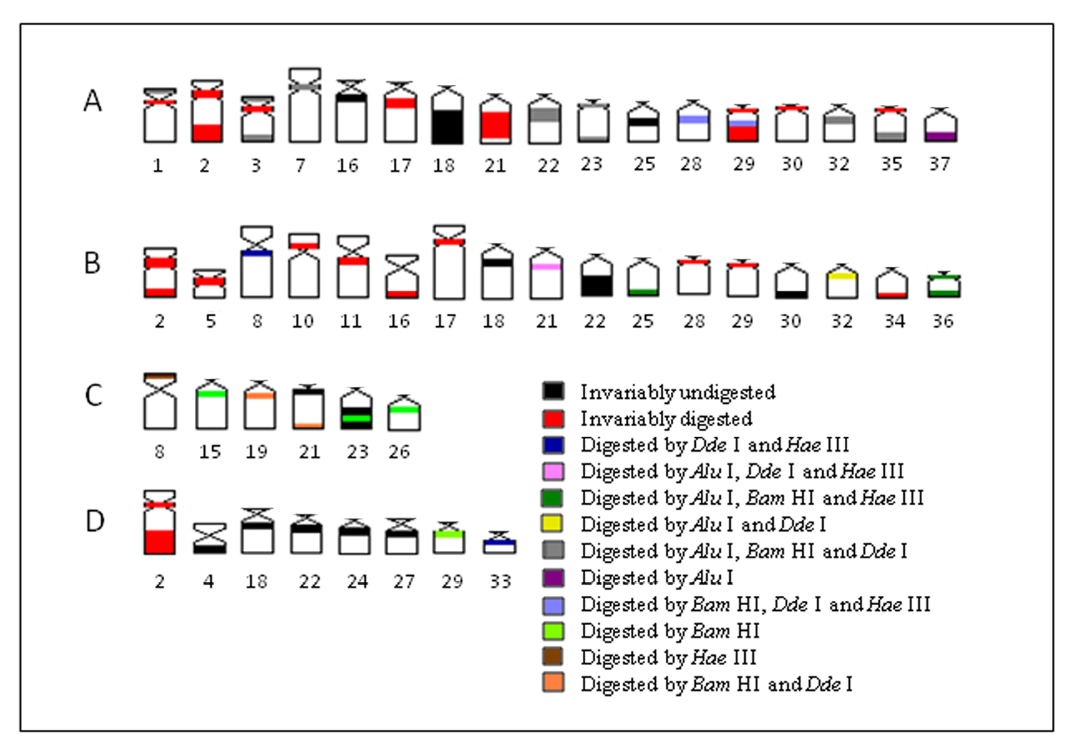
A–D Schematic ideogram of chromosomal pairs from populations A, B, C and D of Hypostomus prope unae, showing the combined banding pattern after digestion using Alu I, Bam HI, Hae III and Dde I.

The present data indicate that some heterochromatin regions in different chromosomes and/or populations share a similar composition, while others would present a unique composition. Thus, the banding pattern observed reflects directly the molecular nature of heterochromatin regions (Sánchez et al. 1991), although a differential access to target sequences by the RE might be present as well.

Such remarkable heterogeneous banding pattern shows that the populations of Hypostomus prope unae bear several heterochromatin families composed of distinct specific types of highly repetitive DNA. A similar finding was reported in the salmonids Salmo salar Linnaeus, 1758(Albuín et al. 1994) and Salmo trutta Linnaeus, 1758 (Sánchez et al. 1991), in which RE digestion resulted in differential heterochromatin digestion in specific chromosomal regions.

According to Schweizer and Loidl (1987), a non-random arrangement of chromosomes during interphase might favor the linkage between certain chromosomal regions and further heterochromatin dispersal to equilocal sites from one chromosome to another, as previously proposed for the distribution of interstitial heterochromatin in other Hypostomus species (Artoni and Bertollo 1999). It seems plausible to infer that those heterochromatin segments sharing a similar composition would have a common origin and have been dispersed to similar chromosomal regions of Hypostomus prope unae. Through their karyoevolutionary history, these segments could have been amplified or accumulated by unequal exchanges, transpositions and/or regional duplications as similarly hypothesized for the marine fish Centropyge aurantonotus Burgess, 1974 (Affonso and Galetti Jr. 2005). Consequently, the chromosomal divergence among the studied populations have possibly been related to rearrangements in the heterochromatin organization and fixed either by genetic drift or by natural selection if some adaptive role is assumed.

Although inter-population differences were detected by both C-banding and RE digestion, some heterochromatin regions remained resistant to enzymatic digestion among populations, mainly in population D, revealing a higher differentiation in the DNA composition and/or heterochromatin organization in the latter. This population is also more divergent than the others because of its high frequency of interstitial C bands instead of terminal ones (Figs 5, 6).

Differences in heterochromatin patterns have been commonly reported in Neotropical fishes, including species from northeastern coastal basins (Jacobina et al. 2009). However, evolutionary mechanisms of heterochromatin differentiation among fish populations are usually related to polymorphic conditions being rarely detected within a single basin (Molina et al. 2008). Thus, the present results indicate that gene flow among Hypostomus prope unae along Contas river basin is absent, favoring the fixation of divergent heterochromatin patterns.

It should be pointed out that the nucleolar organizer regions (2nd pair) was digested by all tested enzymes independently on the population, demonstrating that the distinct target sequences are “concertedly” interspersed along this region, even when NOR-associated heterochromatin was not detected, as observed in population C. Such behavior differs from the pattern observed by Sanches et al. (1990) that reported a differential NOR digestion indicative of a high amount of target sequences for Dde I and Hae III but a moderate number of restriction sites for Alu I.

Moreover, heteromorphic segments were observed between some chromosomal pairs in populations A (pairs 18, 21 and 37) and B (pair 22). Nonetheless, only the 21st pair in population A presented the target sequences for the selected RE, while the other heteromorphic segments proved to be resistant to their digestion activity.

Reports about restriction enzymes in Neotropical fish cytogenetics are scarce what hinders a detailed comparative analysis. However, this approach seems to be highly informative for species characterized by large amounts of heterochromatin as that presently studied, being able to reveal several genomic particularities. Moreover, repetitive DNA sequences might provide efficient chromosomal markers useful for evolutionary studies, identification of chromosomal rearrangements and sex differentiation (Ferreira and Martins 2008).

As commonly reported in fishes of the genus Hypostomus (e.g., Milhomem et al. 2010), the present cytogenetic analyses were able to differentiate the four studied populations of Hypostomus prope unae, thereby reinforcing their evolutionary divergence along Contas river basin and their cryptic species diversity.

Acknowledgements

The authors would like to thank professors Ana Maria Waldschmidt (UESB) for providing the conditions and part of the material used in the present research, Cláudio Henrique Zawadzki (UEM-NUPELIA) for identifying the specimens, Antonio Carlos Bertollo (UFSCar) and André Laforga Vanzella (UEL) for their helpful comments and Conselho Nacional de Desenvolvimento Científico e Tecnológico (CNPq) for the financial support.

References
Affonso PRAM, Galetti Jr PM (2005) Chromosomal diversification of reef fishes from genus Centropyge (Perciformes, Pomacanthidae). Genetica 123 (3): 227-233. doi: 10.1007/s10709-004-3214-x
Albuín M, Amaro R, Sanchez L (1994) Improving Salmo salar karyotype: restriction enzyme and replication banding. Cytobios 78: 143-152.
Artoni RF, Venere PC, Bertollo LAC (1998) A heteromorphic ZZ/ZW sex chromosome system in fish, genus Hypostomus (Loricariidae). Cytologia 63: 421-425.
Artoni RF, Bertollo LAC (1999) Nature and distribution of constitutive heterochromatin in fishes, genus Hypostomus (Loricariidae). Genetica 106 (3): 209-214. doi: 10.1023/A:1003957719178
Bertollo LAC, Takahashi CS, Moreira-Filho O (1978) Cytotaxonomic considerations on Hoplias lacerdae (Pisces, Erythrinidae). Brazilian Journal of Genetics 1: 103-120.
Bianchi MS, Bianchi NO, Pantelias GE, Wolff S (1985) The mechanism and pattern of banding induced by restriction endonucleases in human chromosomes. Chromosoma 91 (2): 131-136.
Bitencourt JA (2010) Análise citogenética de espécies do gênero Hypostomus (Loricariidae; Hypostominae) das bacias do rio de Contas e Recôncavo Sul/Bahia. Mastership Dissertation, Londrina, PR.: Universidade Estadual de Londrina. [In Portuguese]
Bouza C, Sanchez L, Martínez P (1994) karotypic characterization of turbot (Scophthalmus maximus) with conventional, fluorochrome and restriction endonuclease-banding techniques. Marine Biology 120 (4): 609-613. doi: 10.1007/BF00350082
Burkholder GD, Weaver MG (1977) DNA-protein interactions and chromosome banding. Experimental Cell Research 106 (2): 251-262. doi: 10.1016/0014-4827(77)90290-7
Carvalho RA, Dias AL (2005) Karyotypic characterization of Iheringichthys labrosus (Pisces, Pimelodidae): C-, G- and restriction endonuclease banding. Genetics and Molecular Research 4 (4): 663-667.
Cau A, Salvadori S, Deiana AM, Bella JL, Mezzanotte R (1988) The characterization of Muraena helena L. mitotic chromosomes: karyotype, C-banding, nucleolar organizer regions, and in situ digestion with restriction endonucleases. Cytogenetics and Cell Genetics 47 (4): 223-226. doi: 10.1159/000132554
Cereali SS, Pomini E, Rosa R, Zawadzki CH, Froehlich O, Giuliano-Caetano L (2008) Karyotype description of two species of Hypostomus (Siluriformes, Loricariidae) of the Planalto da Bodoquena, Brazil. Genetics and Molecular Research 7 (3): 583-591.
Ferreira IA, Martins C (2008) Physical chromosome mapping of repetitive DNA sequences in Nile tilapia Oreochromis niloticus: evidences for a differential distribution of repetitive elements in the sex chromosomes. Micron 39 (4): 411-418. doi: 10.1016/j.micron.2007.02.010
Gosálvez J, Bella JL, López-Fernández C, Mezzanotte R (1986) Correlation between constitutive heterochromatin and restriction enzyme resistant chromatin in Arcyptera tornosi (Orthoptera). Heredity 59: 173-180. doi: 10.1038/hdy.1987.112
Jacobina UP, Affonso PRAM, Carneiro PLS, Dergam JA (2009) Biogeography and comparative cytogenetics between two populations of Hoplias malabaricus (Bloch, 1794) (Ostariophysi, Erythrinidae) from coastal basins in the State of Bahia, Brazil. Neotropical Ichthyology 7 (4): 617-622. doi: 10.1590/S1679-62252009000400009
Juan C, Gosálvez J, Petitpierre E (1990) Improving beetle karyotype analysis: restriction endonuclease banding of Tenebrio molitor chromosomes. Heredity 65: 157-162. doi: 10.1038/hdy.1990.83
Kaelbling M, Miller DA, Miller OJ (1984) Restriction enzyme banding of mouse metaphase chromosomes. Chromosoma 90 (2): 128-132.
Kantek DLZ, Noleto RB, Fenocchio AS, Cestari MM (2007) Cytotaxonomy, heterochromatic polymorphism and natural triploidy of a species of Astyanax (Pisces, Characidae) endemic to the Iguaçu River Basin. Brazilian Archives ofBiology and Technology 50 (1): 67-74. doi: 10.1590/S1516-89132007000100008
Kavalco KF, Pazza R, Bertollo LAC, Moreira-Filho O (2004) Heterochromatin characterization of four fish species of the family Loricariidae (Siluriformes). Hereditas 141 (3): 237-242. doi: 10.1111/j.1601-5223.2004.01850.x
Kavalco KF, Pazza R, Bertollo LAC, Moreira-Filho O (2005) Karyotypic diversity and evolution of Loricariidae (Pisces, Siluriformes). Heredity 94: 180-186. doi: 10.1038/sj.hdy.6800595
Lee MR, Elder FFB (1980) Yeast stimulation of bone marrow mitosis for cytogenetic investigations. Cytogenetics and Cell Genetics 26: 36-40. doi: 10.1159/000131419
Levan A, Fredga K, Sandberg AA (1964) A nomenclature for centromeric position on chromosomes. Hereditas 52: 201-220. doi: 10.1111/j.1601-5223.1964.tb01953.x
Lima-de-Faria A, Isaksson M, Olsson E (1980) Action of restriction endonucleases on the DNA and chromosomes of Muntiacus muntjak. Hereditas 92: 267-273. doi: 10.1111/j.1601-5223.1980.tb01707.x
Lloyd MA, Thorgaard GH (1988) Restriction endonuclease banding of rainbow trout chromosomes. Chromosoma 96 (2): 171-177. doi: 10.1007/BF00331049
Lorite P, Garcia MF, Carrillo JA, Palomeque T (1999) Restriction endonuclease chromosome banding in Tapinoma nigerrimum (Hymenoptera, Formicidae). Hereditas 131: 197-201. doi: 10.1111/j.1601-5223.1999.00197.x
Lozano R, Ruiz Rejon C, Ruiz Rejon M (1991) An analysis of coho salmon chromatin by means of C-banding, AG- and fluorochrome staining, and in situ digestion with restriction endonucleases. Heredity 66: 403-409. doi: 10.1038/hdy.1991.50
Maistro EL, Foresti F, Oliveira C (1999) R- and G-band patterns in Astyanax scabripinnis paranae (Pisces, Characiformes, Characidae). Genetics and Molecular Biology 22 (2): 201-204. doi: 10.1590/S1415-47571999000200011
Maistro EL, Oliveira C, Foresti F (2000) Cytogenetic analysis of A and B chromosomes of Prochilodus lineatus (Teleostei, Prochilodontidae) using different restriction enzyme banding and staining methods. Genetica 108 (2): 119-125. doi: 10.1023/A:1004063031965
Marchi A, Mezzanotte R (1988) Restriction endonuclease digestion and chromosome banding in the mosquito, Culiseta longiareolata (Diptera: Culicidae). Heredity 60: 21-26. doi: 10.1038/hdy.1988.4
Marchi A, Mezzanotte R (1990) Inter- and intraspecific heterochromatin variation detected by restriction endonuclease digestion in two sibling species of the Anopheles maculipennis complex. Heredity 65: 135-142. doi: 10.1038/hdy.1990.80
Mezzanotte R, Bianchi U, Vanni R, Ferruci L (1983) Chromatin organization and restriction nuclease activity on human methaphase chromosomes. Cytogenetics and Cell Genetics 36 (3): 562-566. doi: 10.1159/000131973
Mezzanote R, Ferrucci L, Vanni R, Sumner AT (1985) Some factors affecting the action of restriction endonucleases on human metaphase chromosomes. Experimental Cell Research 161 (1): 247-253. doi: 10.1016/0014-4827(85)90509-9
Miller DA, Tantravahi R, Dev VG, Miller OJ (1976) Q- and C-band chromosome markers in inbred strains of Mus musculus. Genetics 81 (1): 67-75.
Miller DA, Choi JC, Miller JO (1983) Chromosome localization of highly repetitive human DNAs and amplified ribosomal DNA with restriction enzymes. Science 219 (4583): 395-397. doi: 10.1126/science.6294832
Milhomem SSR, Castro RR, Nagamachi CY, Souza ACP, Feldberg E, Pieczarka JC (2010) Different cytotypes in fishes of the genus Hypostomus Lacépède, 1803 (Siluriformes: Loricariidae) from Xingu river (Amazon region, Brazil. Comparative cytogenetics 4 (1): 45-54.
Molina WF (2001) An alternative method for mitotic stimulation in fish cytogenetics. Chromosome Science 5 (3): 149-152.
Molina WF, Shibatta O, Galetti Jr PM (2008) Chromosomal evidence of population subdivision in the freshwater fish Leporinus elongatus in the Upper Paraná River basin. Genetics and Molecular Biology 31 (1): 270-274. doi: 10.1590/S1415-47572008000200020
Pieczarka JC, Nagamachi CY, Barros RMS, Mattevi MS (1996) Analysis of constitutive heterochromatin by fluorochromes and in situ digestion with restriction enzymes in species of the group Callithrix argentata (Callitrichidae, Primates). Cytogenetics and Cell Genetics 72 (4): 325-330. doi: 10.1159/000134215
Rubert M, Zawadzki CH, Giuliano-Caetano L (2008) Cytogenetic characterization of Hypostomus nigromaculatus (Siluriformes: Loricariidae). Neotropical Ichthyology 6 (1): 93-100. doi: 10.1590/S1679-62252008000100011
Sánchez L, Martínez P, Viñas A, Bouza C (1990) Analysis of the structure and variability of nucleolar organizer regions of Salmo trutta by C-, Ag-, and restriction endonuclease banding. Cytogenetics and Cell Genetics 54(1–2): 6-9. doi: 10.1159/000132944
Sánchez L, Martínez P, Bouza C, Viñas A (1991) Chromosomal heterochromatin differentiation in Salmo trutta with restriction enzymes. Heredity 66: 241-249. doi: 10.1038/hdy.1991.30
Sumner AT (1972) A simple technique for demonstrating centromeric heterochromatin. Experimental Cell Research 75: 304-306. doi: 10.1016/0014-4827(72)90558-7
Schweizer D, Loidl J (1987) A model for heterochromatin dispersion and the evolution of C-bands patterns. Chromosome Today 9: 61-74.
Swarça AC, Fenocchio AS, Cestari MM, Dias AL (2005) First chromosome data on Steindachneridion scripta (Pisces, Siluriformes, Pimelodidae) from Brazilian rivers: Giemsa, CBG, G, and RE banding. Genetics and Molecular Research 4 (4): 734-741.